Animal Cell Labeled Microtubules / Unique Characteristics of Eukaryotic Cells | Microbiology - Microfilaments are organelle cells formed from actin and myosin proteins.
byJunko Walch-
0
Animal Cell Labeled Microtubules / Unique Characteristics of Eukaryotic Cells | Microbiology - Microfilaments are organelle cells formed from actin and myosin proteins.. These straight, hollow cylinders are found throughout the cytoplasm of all. The cell is the basic unit of life. The outer diameter of a microtubule is between 23 and 27 nm while the inner diameter is between 11. Both of these structures are found in animal cells, but not plant cells. Essentially, the two aggregated centrioles act as anchors for the rest of the cell's.
Microtubules are exactly how they sound: An animal cell ranges in size from 10 to 30 µm. Essentially, the two aggregated centrioles act as anchors for the rest of the cell's. Microtubules in the cell consist of microscopic structures formed in hollow tubes and constructed in a series of linear rings. Animal cells are common names for eukaryotic cells that make up animal tissue.
Jello Animal Cell Craft - Enchanted Learning Software from www.zoomschool.com There are three microtubules in each group. Under the microscope, an animal cell shows many different parts called organelles, that work together to keep. Microtubules in the cell consist of microscopic structures formed in hollow tubes and constructed in a series of linear rings. Essentially, the two aggregated centrioles act as anchors for the rest of the cell's. Our body starts its existence at fertilization from a single cell, the diploid zygote. Microtubules are slender, cylindrical filaments found in the cytoskeleton of plant and animal cells. The cells of plants and fungi do not have centrosomes, and instead the nuclear envelope—the membrane surrounding the cell's. Animal cells are the types of cells that make up most of the tissue cells in animals.
The cell is the basic unit of life.
In animal cells, microtubules radiate outwards from an organelle in the center of the cell called a centrosome, which is a microtubule organizing center (mtoc). Animal cells are common names for eukaryotic cells that make up animal tissue. The centrosomes is where microtubules are made. Microfilaments are organelle cells formed from actin and myosin proteins. Microscopic hollow tubes found inside eukaryotic cells and some prokaryotic bacteria cells that provide structure and motor functions. They are composed of the protein tubulin and are influenced by tubulin modulators. The cytoplasmic microtubules in animal cells are connected with the satellites of the centrioles and are similar to the mitotic spindle fibers. Animal cells are unique in that they contain special organelles for the construction and maintenance of microtubules, organelles known as centrioles. All organisms are made up of cells (or in some cases, a single cell). An animal cell ranges in size from 10 to 30 µm. Microtubules as their name implies, microtubules are small hollow tubes. Most of the cells size range between 1 illustrated in figure 2 is a pair of fibroblast deer skin cells that have been labeled with fluorescent probes microtubules. These hollow rods help give structure and shape to the cell.
Our body starts its existence at fertilization from a single cell, the diploid zygote. The cell may contain a pair of centrioles (or microtubule organizing centers in plants) both of which are organizational sites for microtubules. These hollow rods help give structure and shape to the cell. Microtubules as their name implies, microtubules are small hollow tubes. The walls of the microtubule are made of with a diameter of about 25 nm, microtubules are the widest components of the cytoskeleton.
Animal Cells and the Membrane-Bound Nucleus from www.thoughtco.com Cell animal vector prokaryotic biology illustration multicellular nucleus ribosome structure unicellular book cell biology centrioles chromosomes cytoplasm dna endoplasmic reticulum ethics eukaryotic functions fundamental genetic golgi apparatus health labels laboratory lysosome medical membrane. Microtubules in the cell consist of microscopic structures formed in hollow tubes and constructed in a series of linear rings. The outer diameter of a microtubule is between 23 and 27 nm while the inner diameter is between 11. Microtubules are polymers of tubulin that form part of the cytoskeleton and provide structure and shape to eukaryotic cells. Microfilaments bear a resemblance to microtubules but microfilaments are softer and smaller in diameter. The cellular organization of microtubules varies between cell types, but in most cells, the minus ends of microtubules are anchored to the centrosomes near the nucleus while the plus ends radiate towards the periphery of the cell. There are three microtubules in each group. In animal cells, microtubules radiate outwards from an organelle in the center of the cell called a centrosome, which is a microtubule organizing center (mtoc).
The centrosomes is where microtubules are made.
The cellular organization of microtubules varies between cell types, but in most cells, the minus ends of microtubules are anchored to the centrosomes near the nucleus while the plus ends radiate towards the periphery of the cell. Also cilia and flagella are made of microtubules. In the complete animal cell centrosome, the two centrioles are arranged such that one is perpendicular to. Microtubules involved in movement of the cell (cilia and flagella and the basal bodies that connect them to the cell) very stable and not dynamic like the. Cell animal vector prokaryotic biology illustration multicellular nucleus ribosome structure unicellular book cell biology centrioles chromosomes cytoplasm dna endoplasmic reticulum ethics eukaryotic functions fundamental genetic golgi apparatus health labels laboratory lysosome medical membrane. Animal cells are common names for eukaryotic cells that make up animal tissue. Microtubules allow motor proteins like kinesin and dynein to carry vesicles (packages of stuff that will be delivered to a different place in the cell). Different kinds of animals have microtubules: Microtubules in the cell consist of microscopic structures formed in hollow tubes and constructed in a series of linear rings. The cytoplasmic microtubules in animal cells are connected with the satellites of the centrioles and are similar to the mitotic spindle fibers. The walls of the microtubule are made of with a diameter of about 25 nm, microtubules are the widest components of the cytoskeleton. Conduit pt et al., centrosome function and assembly in animal cells. Microfilaments bear a resemblance to microtubules but microfilaments are softer and smaller in diameter.
Organization and shape of animal cells, intracellular organelle mobility, maintain axons, spindle formation. Microtubules can grow as long as 50 micrometres and are highly dynamic. Most of the cells size range between 1 illustrated in figure 2 is a pair of fibroblast deer skin cells that have been labeled with fluorescent probes microtubules. Microtubules as their name implies, microtubules are small hollow tubes. Cell animal vector prokaryotic biology illustration multicellular nucleus ribosome structure unicellular book cell biology centrioles chromosomes cytoplasm dna endoplasmic reticulum ethics eukaryotic functions fundamental genetic golgi apparatus health labels laboratory lysosome medical membrane.
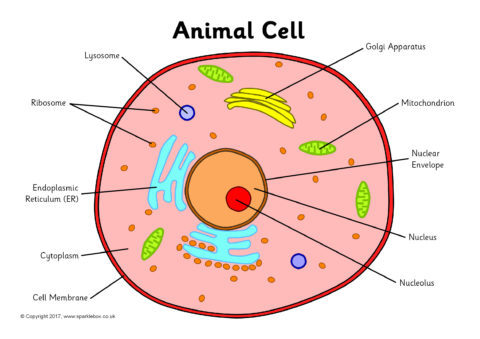
Preview from www.sparklebox.co.uk Microtubules are polymers of tubulin that form part of the cytoskeleton and provide structure and shape to eukaryotic cells. One of the primary concerns with using fluorescent taxanes to label microtubules is the possibility of cytotoxicity upon continuous incubation. Animal cells from the basic structural units of all tissues and organs of the body. Microtubules are exactly how they sound: Cilia are projections from a cell that can m. Microscopic hollow tubes found inside eukaryotic cells and some prokaryotic bacteria cells that provide structure and motor functions. Each centriole is a ring of nine groups of fused microtubules. Animal cells are the types of cells that make up most of the tissue cells in animals.
Also cilia and flagella are made of microtubules.
Two or more centrioles will aggregate perpendicularly to each other. Cilia are projections from a cell that can m. The centrosomes is where microtubules are made. Microtubules can grow as long as 50 micrometres and are highly dynamic. The cellular organization of microtubules varies between cell types, but in most cells, the minus ends of microtubules are anchored to the centrosomes near the nucleus while the plus ends radiate towards the periphery of the cell. Animal cells are unique in that they contain special organelles for the construction and maintenance of microtubules, organelles known as centrioles. The cell is the basic unit of life. Microtubules in the cell consist of microscopic structures formed in hollow tubes and constructed in a series of linear rings. Animal cells are of various sizes and have irregular shapes. Animal cells from the basic structural units of all tissues and organs of the body. Microfilaments are organelle cells formed from actin and myosin proteins. They are composed of the protein tubulin and are influenced by tubulin modulators. The cells of plants and fungi do not have centrosomes, and instead the nuclear envelope—the membrane surrounding the cell's.

:max_bytes(150000):strip_icc()/Eukaryotic_Cell_animal-59df7d9f03f40200104fcd0b.jpg)